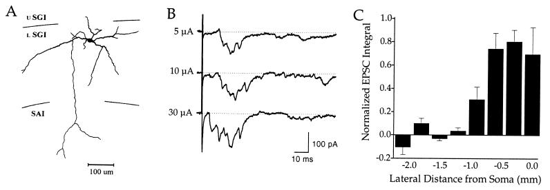
Figure 5.
Synaptic responses of a cell in the lower intermediate gray layer. (A) The axon of this neuron descends to exit the colliculus through its deeper layers. (B) Prolonged bursts of EPSCs evoked after single stimuli of different intensities. (C) Relationship between postsynaptic responses and stimulating electrode position. This cell was located below the last electrode in the array (i.e., 0.0 mm). For definitions of abbreviations used in A, see the Fig. 1 legend.

