Figure 3.
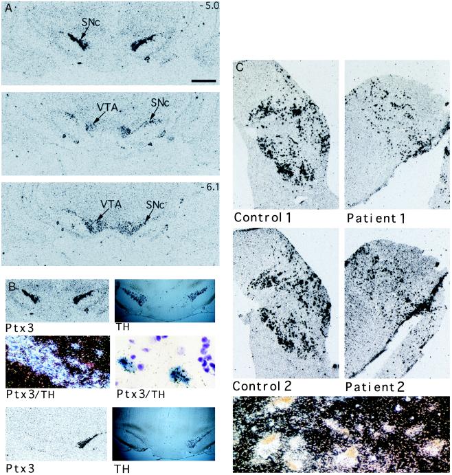
Expression of Ptx3 in the mesencephalic dopaminergic system of rat and human. (A) The expression pattern of Ptx3 in rat brain starting anterior at the substantia nigra compacta (SNc) to posterior at the ventral tegmental area (VTA). The size bar (Top) represents a distance of 1 mm. The rostral–caudal positions of the Top and Bottom are indicated as millimeters relative to bregma in the upper right corner. (B) Double in situ hybridizations using a DIG-labeled tyrosine hydroxylase (TH) cRNA probe and a 35S-labeled Ptx3 cRNA probe show a similar hybridization pattern for both probes in the tegmentum of the rat brain (Top Left and Top Right). Microscopic examination (Middle) shows TH-positive cells in blue and Ptx3 labeled by silver grains (Middle Left, dark-field image; Middle Right, higher magnification, bright-field image). A complete overlap is found. In situ hybridization for TH and Ptx3 expression of animals with a unilateral lesion of dopaminergic neurons within the mesDA system. (Lower) The animals displayed increased turning behavior in the direction of the lesioned side. Both TH and Ptx3 expression was lost at the lesioned side. (C) Ptx3 expression in the human substantia nigra of two healthy controls (Control 1 and 2) and two Parkinson patients (Patient 1 and 2). The sections of the patients show a lower density of Ptx3-expressing cells, which correlates with the loss of dopaminergic neurons within this region. (Lower) A dark-field image showing Ptx3 expression (silver grains) colocalizing with typical brown-pigmented dopaminergic neurons of the human substantia nigra. Material was obtained from the Netherlands Brain Bank. Patient material was diagnosed clinically and pathologically.

