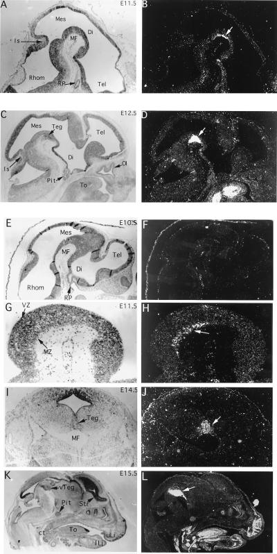
Figure 4.
In situ hybridization of Ptx3 in embryonic mouse brain sections between E10.5 and E15.5. Micrographs of bright-field and dark-field images of the same sections are shown. (A and B) At E11.5, expression is seen in a layer of postmitotic cells (arrow) in the mesencephalon (Mes) lining the mesencephalic flexure (MF). (C and D) At E12.5 the layer of expressing cells has thickened as part of the developing tegmentum (Teg). Extraneural expression is seen in the tongue (To) but not in the developing pituitary (Pit). (E and F) Expression of Ptx3 was not detected in the brain at E10.5. Lack of expression in Rathke’s pouch (RP), a site of Ptx1 and Ptx2 expression (11–14), confirms the specificity of the hybridization reaction. (G and H) Higher magnification of the tegmental region expressing Ptx3 at E11.5. Expression is detected ventrally in the marginal zone (MZ) of the neuroepithelium. (I and J) A coronal section of an E14.5 mouse brain shows the lateral extent of Ptx3 expression in the developing tegmentum (Teg). (K and L) A sagittal section of an E15.5 mouse head shows strong expression of Ptx3 in the ventral tegmentum (vTeg). In contrast to other hybridizations that were performed with 5′ or 3′ Ptx3 probes, the probe used for this experiment contained the homeodomain, and there is weak cross-reactivity with other members of the Ptx family outside the brain, e.g., the pituitary (Pit) (11–14). Mes, mesencephalon; Di, diencephalon; Tel, Telencephalon; Rhom, rhombencephalon; MF, mesencephalic flexure; RP, Rathke’s pouch; Is, isthmus; Pit, pituitary; To, tongue; Ol, olfactory epithelium; MZ, marginal zone; VZ, ventricular zone; Str, striatum; vTeg, ventral tegmentum; ct, cartilage of the throat; uLi, upper lip, lLi, lower lip.

