
Figure 1. Induction of apoptosis in fatty acid and butyrate-treated colonocytes.
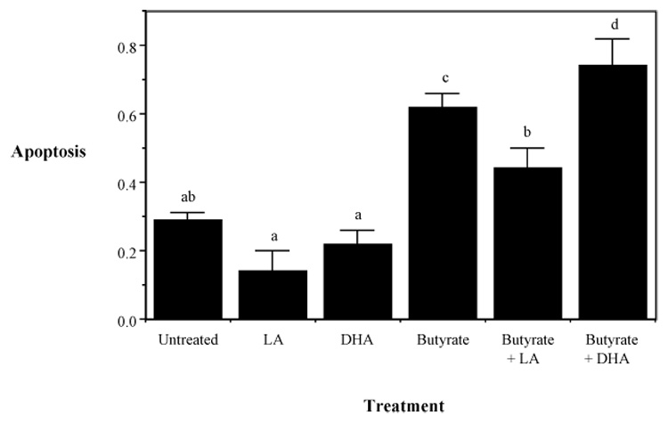
Cultures contained 5 mM butyrate and 50 µM DHA, LA or no fatty acid treatment. Apoptosis was measured by DNA fragmentation ELISA. Data represent mean absorbance at 405 nm ± SE divided by the total number of adherent cells per dish, n=4–6 separate wells from 1 of 4 representative experiments. Values not sharing the same letters are significantly different (P < 0.05). Adapted from Ng et al (2005).