
Fig 4.
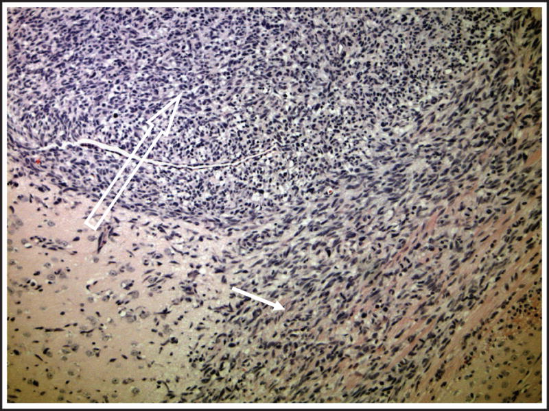
Growth of intracranial tumors established from flank xenografts. A representative section of an orthotopic tumor displays a close resemblance to primary tumors regarding advanced stage of growth. The tumor revealed high cellularity (open arrow) and invasion into corpus callosum (closed arrow).