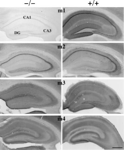
Figure 2.
Light microscopic localization of m1–m4 in the hippocampus of m1 knockout (−/−, Left) and wild-type mice (+/+, Right). Note the absence of m1 immunoreactivity in the hippocampus and overlying cortex from the mutant mice when compared with the dark staining evident in the wild-type mice. Minimal background staining in the m1-deficient mice was similar to controls in which the primary antibody was omitted for mutant and wild-type brain sections. CA1 and CA3, regions of Ammon’s horn; DG, dentate gyrus. (Bar = 500 μm.)

