
Figure 3.
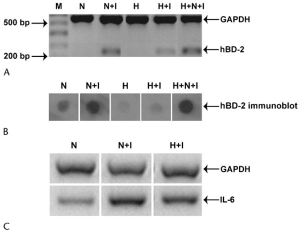
The ability of IL-1β to stimulate hBD-2 and IL-6 expression in HCECs is reduced by 500 mOsm/kg hyperosmolar media. A, SV40-HCECs (n = 4) were cultured for 24 hours in serum-free normal osmolality media (N) or 500 mOsm/kg (NaCl) hyperosmolar media (H). At the end of the incubation, either (1) 10 ng/mL IL-1β was directly added to the culture media of cells in the normal-osmolality media (N+I) and to those in hyperosmolar media (H+I) for 6 hours or (2) the hyperosmolar media was aspirated and substituted with normal-osmolality media containing 10 ng/mL IL-1β for 6 hours (H+N+I). The figure shows representative RT-PCR products for GAPDH and hBD-2 from 1 experiment. M, marker lane. Similar results were obtained with P-HCECs (n = 2, data not shown). B, P-HCECs (n = 2) were cultured for 24 hours in extract-free normal-osmolality media (N) or 500 mOsm/kg (NaCl) hyperosmolar media (H). Experimental conditions were similar to those described for panel A. The figure shows changes in hBD-2 protein secretion by P-HCECs for the various conditions. Similar results were obtained with SV40-HCECs (n = 1, data not shown). C, The experimental conditions were similar to those in panel A. The figure shows RT-PCR products for GAPDH and IL-6. The data are representative of 1 experiment repeated twice with SV40-HCECs. Similar results were obtained with P-HCECs (n = 2, data not shown).