
Figure 1.
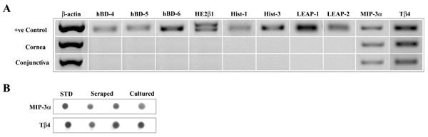
Expression of MIP-3α and Tβ4 mRNA and protein by ocular surface epithelia. (A) RT-PCR. The figure shows representative results for cornea = scraped human corneal epithelium (n = 3); conjunctiva = primary cultured human conjunctival epithelial cells (n = 2); + ve controls = positive controls: testis (hBD 4–6, HE2β1, MIP-3α), salivary gland (Hist-1, -3), liver (LEAP 1-2), and thymus (Tβ4). (B) Immunoblotting. The figure shows representative results for: STD (standard) = 5 ng human rMIP-3α or 10 ng Tβ4 synthetic peptide; scraped = 25 μg cellular protein from two scraped human corneal epithelial samples (n = 2); cultured = 25 μg cellular protein from primary cultured human corneal epithelial cells (n = 3).