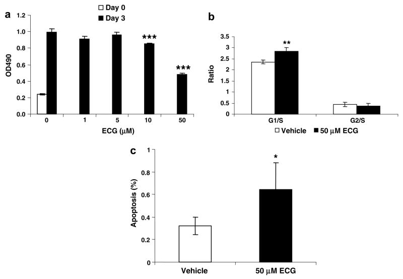
Fig. 4.
Growth retardation and apoptosis of SCC7 cells treated with ECG. (a) Effect of ECG on SCC7 cell growth. SCC7 cells were plated at 1000 cells/well in a 96-well plate and incubated with the vehicle (DMSO) or various concentrations of ECG for 72 h. Cell growth was measured using CellTiter 96 Aqueous One Solution Cell Proliferation Assay (Promega, Madison, WI). Values are expressed as means ± SD of 6 replicates. (b and c) Cell cycle and apoptosis of ECG-treated SCC7 cells. Cells were plated at 1 × 10⁵ cells/well in 6-well plates, incubated with the vehicle or ECG for 72 h, and analyzed for (b) cell cycle analysis and (c) apoptosis as described in Section 2.

