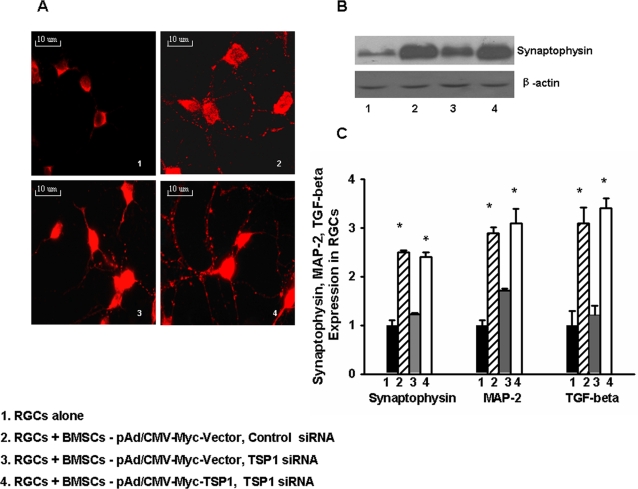
Figure 7. RGC synaptophysin, MAP-2 and TGF-β expression level were determined with and without BMSC (+/− TSP-1 siRNA silencing) co-culturing.
BMSCs were transfected with siRNA oligos and infected with a recombinant adenovirus (pAd/CMV-Myc-TSP-1) expressing TSP-1 before co-culturing as described above. A, After 3 days, immunohistochemical analysis of RGCs for synaptophysin (red dots) shows more synaptic puncta when co-culturing with BMSCs compared to RGCs alone (2 & 4), but fewer when TSP-1 was silenced by siRNA (3). B, Western blot shows synaptophysin in RGCs is up-regulated by TSP-1; (2), which was diminished by TSP-1 silencing (3) and restored by TSP-1-expressing adenoviral rescue (4). C, After 3 days, synaptophysin, MAP-2 and TGF-β expression were determined by real-time RT-PCR (p = 0.019, p = 0.020, p = 0.017, respectively). *p-values≤0.05. (The experiments were repeated at least three times).

