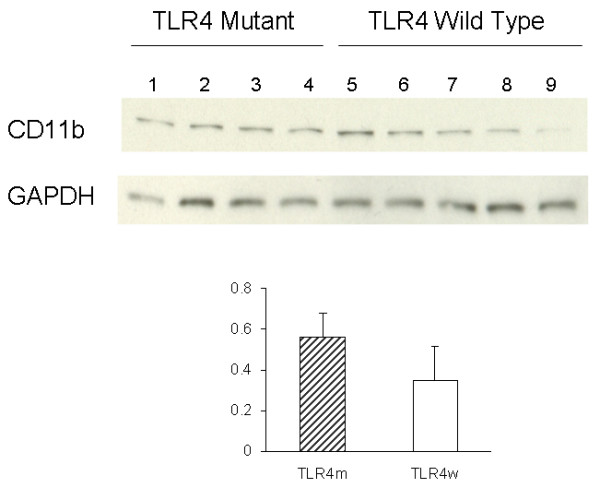
Figure 2.

Levels of CD11b and GAPDH in the cerebral tissue lysates were determined by immunoblotting using anti-CD11b and anti-GAPDH antibodies, respectively. The bar graph represents densitometric quantification of CD11b after normalization with GAPDH (means ± SE). The mean of CD11b levels in TLR4m AD mice is greater than that in TLR4w AD mice but not statistically significant. Lane 1 through 4 are tissue lysates from TLR4m AD mice and lane 5 through 9 are from TLR4w AD mice.
