
Figure 1.
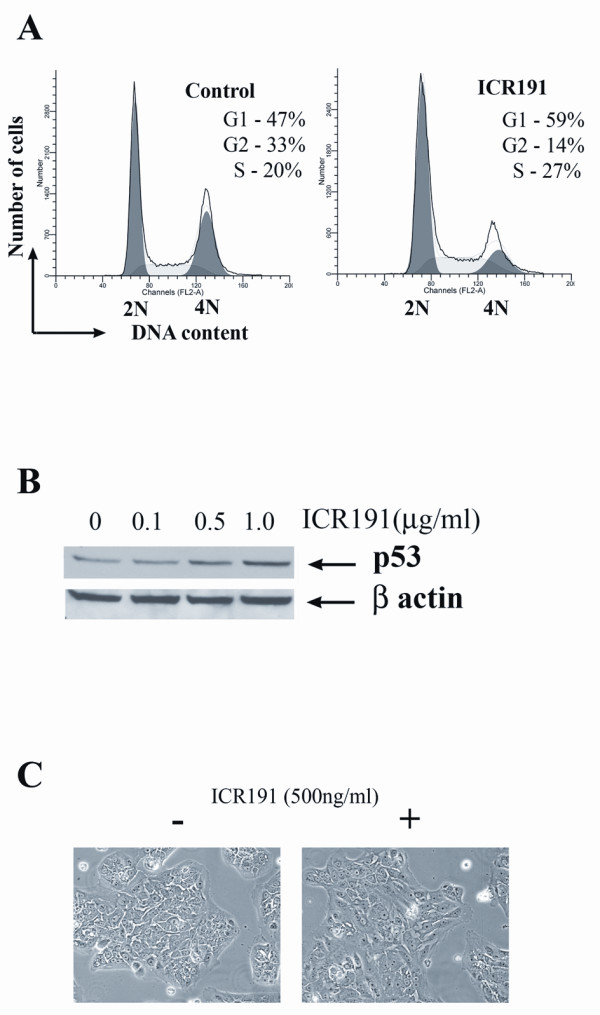
Effect of ICR191 exposure on MCF-10A cells. A. Fluorescence-activated cell-sorting (FACS) analysis demonstrates that MCF-10A cells, after 24 hours of cultivation in a tissue-culture medium containing 500 ng/ml of ICR191, have an increased proportion of cells arrested in the S and G1 phases of the cell cycle. B. Western blot demonstrates the accumulation of P53 protein induced by ICR191 treatment. C. A slightly larger size and flatter appearance of MCF-10A cells after 24 hours of exposure to 500 ng/ml of ICR191.