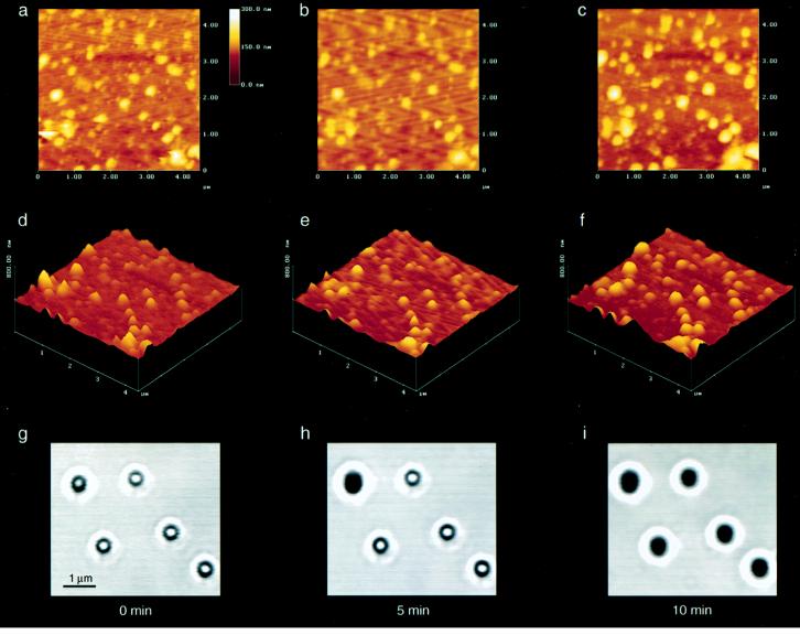
Figure 3.
Increase in size of ZGs in the presence of GTP. (a–c) Two-dimensional AFM images of the same granules after exposure to 20 μM GTP at time 0 (a), 5 minutes (b), and 10 minutes (c). (d–f) The same granules are shown in three-dimensions: the three-dimensional image of the granules at time 0, 5 minutes, and 10 minutes, respectively, after exposure to GTP. (g-i) The GTP-induced increase in size of another group of ZGs observed by confocal microscopy. Confocal images of the same ZGs at time 0, 5 minutes, and 10 minutes after GTP exposure are shown. (Bar = 1 μm.) Values represent one of three representative experiments.

