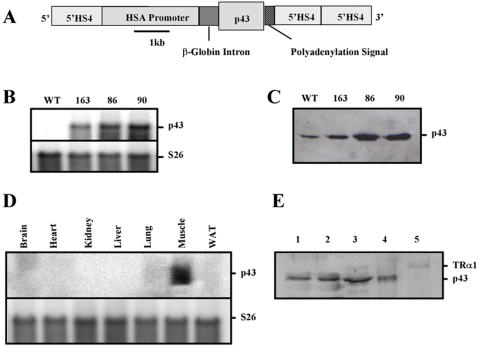
Figure 1. Transgenic expression of p43 in mouse skeletal muscle.
(A) Schematic representation of the construct used for microinjection of [C57BL/6 x CBA] F1 fertilized oocytes. HSA: human α-skeletal actin; 5′HS4: chicken β-globin insulator. (B) mRNAs were isolated from quadriceps from transgenic mice of the 86, 90 and 163 lines versus wild-type (WT) animals and subjected to hybridization analysis with probes for p43. Hybridization with ribosomal S26 probes served as loading control. 20 µg of total RNAs were analyzed. (C) p43 protein levels in quadriceps muscle mitochondria from transgenic mice of the 86, 90 and 163 lines versus wild-type animals, visualized by western-blot using an antibody raised against TRα. 50 µg of mitochondrial proteins were analyzed. (D) mRNAs were isolated from various tissues from transgenic mice of the 86, 90 and 163 lines versus wild-type animals and subjected to hybridization analysis with probes for p43. Hybridization with ribosomal S26 probes served as loading control. 20 µg of total RNAs were analyzed. (E) Western blot of the fractions collected during the nuclear isolation using an antibody raised against TRα. Fractions are whole muscle homogenate-I (1), whole muscle homogenate-II (2), crude nuclear fraction (3), plasma membranes, mitochondria (4), nuclei (5). 25 µg of proteins were analyzed. Arrows indicate the nuclear receptor TRα1 and p43.

