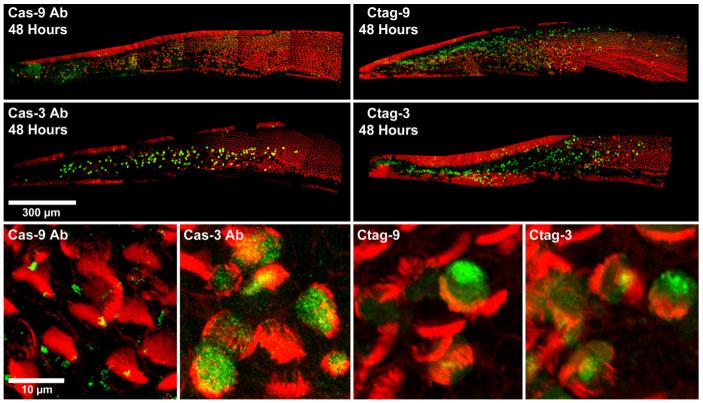
Fig. 1.
Confocal images of cochleae labeled for either activated caspase-9 (green) or activated caspase-3 (green) and phalloidin (red) 48 h AI. Both the antibody (left) and the CaspaTag kit (right) reliably label caspase-9 and caspase-3 positive cells. Scale bar = 300 μm. High magnification images of caspase-9 or caspase-3 positive cells labeled with caspase antibodies or CaspaTag can be seen in the bottom row. For both detection methods, the caspase-9 label appears to be weaker than the caspase-3 label. Scale bar = 10 μm.

