
Fig. 4.
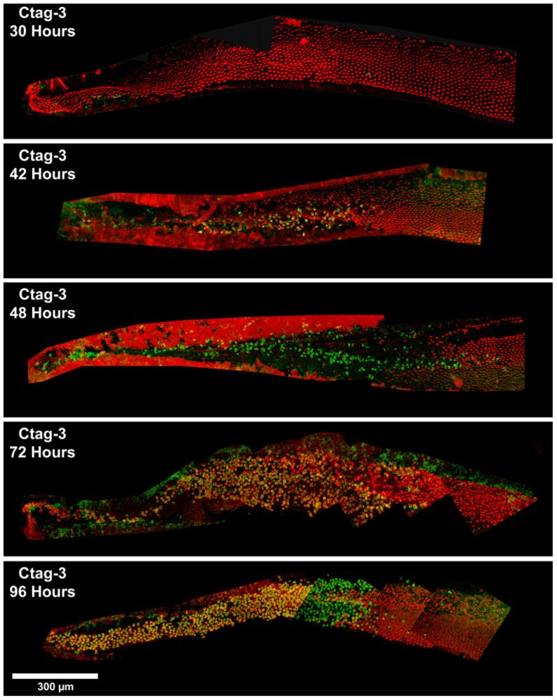
Confocal images of cochleae labeled for activated caspase-3 (green) using a CaspaTag in situ assay kit 30, 42, 48, 72, and 96 h AI. Early time points (30, 42, and 48 h) are co-labeled with phalloidin (red); later time points (72 and 96 h) are co-labeled with myosin VI (red). Overlap of the two labels appears in yellow. The timing of caspase-3 activation using CaspaTag mirrors that seen with caspase-directed antibodies, with labeling first occurring between 30 and 42 h AI. However, the CaspaTag assay appears to label more cells at the later time points, such as 72 and 96 h. Scale bar = 300 μm.