Figure 4.
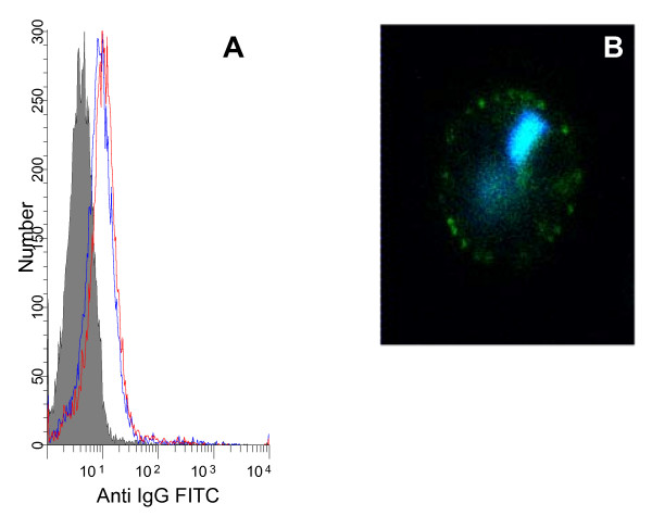
Anti-P2 antibodies react with native VAR2CSA expressed on the surface of infected erythrocytes. The histogram (A) shows staining of red blood cells infected with late stages of NF54var2csa. The IE reacted with rabbit affinity purified antibodies against P2c peptide (red) and DBL5 (blue). The rabbit prebleed is shown in solid grey. The picture (B) shows an IFA image of IE double stained with anti-P2c antibodies (green) and DNA (DAPI) staining (blue).

