
Figure 3. Gating strategy for isolation of BM-derived VSELs by FACS.
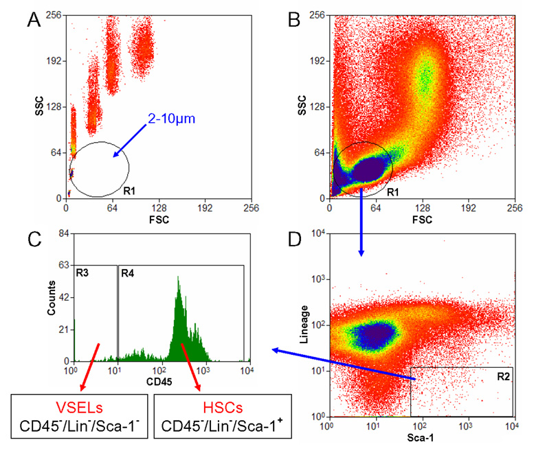
Murine BM-derived VSELs were isolated from full population of bone marrow cells stained for Sca-1, CD45 and hematopoietic lineages markers (Lin) by MoFlo cell sorter (Dako, Carpintera, Ca, USA). Agranular, small events ranging from 2 – 10 µm were included into gate R1 after comparison with six differently sized beads particles with standard diameters of 1, 2, 4, 6, 10 and 15 µm (Flow Cytometry Size beads, Invitrogen; Molecular Probes, Carlsbad, Ca, USA) (Panel A). Cells were visualized by dot plot showing FSC (forward scatter) vs. SSC (side scatter) characteristics, which are related to the size and granularity/complexity of the cell, respectively (Panel B). Objects from region R1 were further analyzed for Sca-1 and Lin expression and only Sca-1+/Lin− events were included into region R2 (Panel D). Population from the region R2 was subsequently sorted based on CD45 marker expression into Sca-1+/Lin−/CD45− VSELs and Sca-1+/Lin−/CD45+ HSCs which are visualized on histogram (Panel C, regions R3 and R4, respectively).