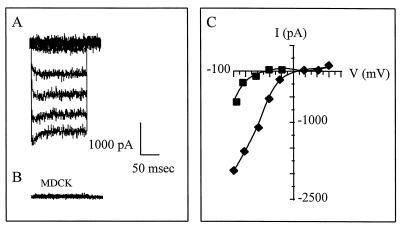
Figure 2.
Functional analysis of epitope-tagged IRK3 channels in MDCK cells. Typical whole cell currents recorded on VSV-IRK3-MDCK cells (A) and mock-transfected MDCK cells (B). The cells were bathed with high K+ solution and voltage steps (200 msec) ranging from −100 to +80 mV were applied from a membrane holding potential of 0 mV. Similar whole cell currents were recorded on AU1- and VSV(Y2F)-IRK3-MDCK cells. (C) Current–voltage (I/V) plot of the whole cell currents recorded with high (140 mM) and low (20 mM) external K in VSV-IRK3-MDCK cells.

