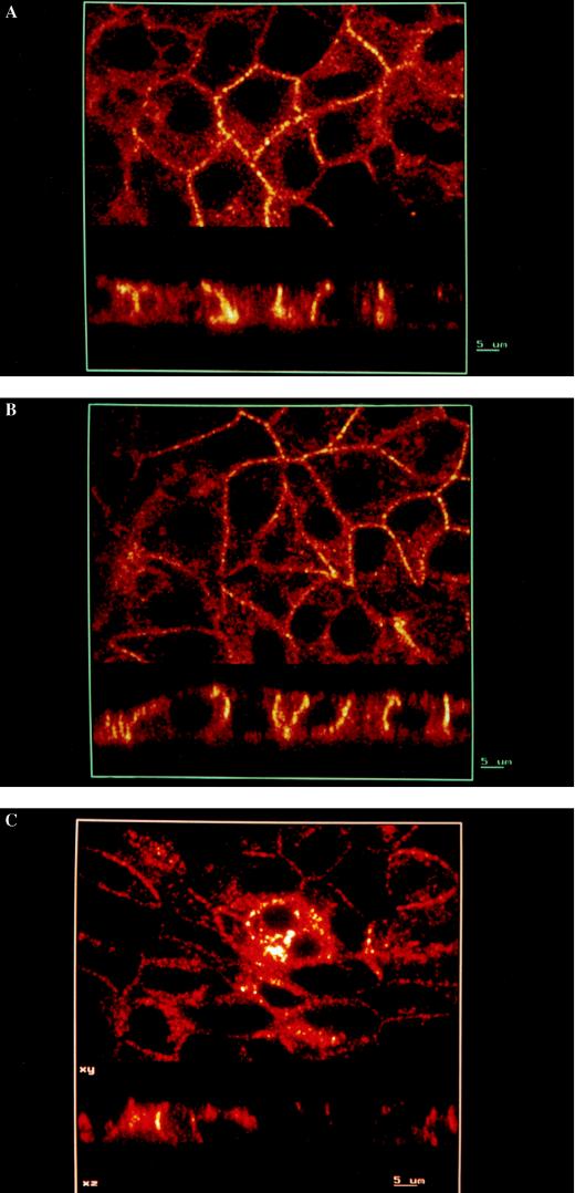
Figure 4.
Basolateral targeting of epitope-tagged IRK3 channels in MDCK cells. Immunolocalization of VSV, VSV(Y2F), and AU1-IRK3 channels in MDCK cells (A–C, respectively). The cells were grown to confluence on permeable supports and processed for immunofluorescence confocal microscopy. In each micrograph, Upper and Lower show horizontal and vertical sections through the cell layer, respectively. The yellow labeling of the basolateral cell membranes indicates specific targeting of the channel proteins to this membrane domain. (Bars = 5 μm.)

