
Figure 3. Electron microscopical analyses of CEC infected with v20US3*K220A.
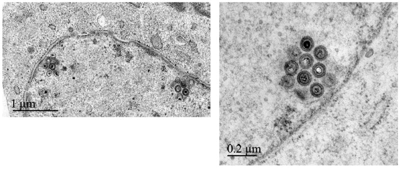
Cells were infected and fixed 3 days later. The v20US3*K220A kinase-negative mutant exhibited the same phenotype as the pUS3 null virus v20ΔUS3 (Schumacher et al., 2005). Primarily enveloped virions accumulated in the perinuclear space forming characteristic invaginations of the inner leaflet of the nuclear membrane. Bars represent 1 mm (left overview panel ) or 200 nm (right panel).