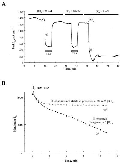
Figure 2.
Extracellular K+ ions protect K channels against the irreversible effect of TEA+. (A) Time course of maximum IK amplitudes after voltage jumps to 40 mV. The axon was bathed in a solution containing 20 mM K+ ions and perfused with 1 mM TEA+ (open bar) for 5 min. IK was reduced to ≈40% of its peak by TEA+ and almost fully recovered when TEA+ was washed out. IK also fully recovered from TEA+ when the extracellular solution contained 10 mM K+. When no K+ was added to the extracellular solution, however, less than 20% of the current recovered after exposure of the axon to TEA+. Experiment SE136A. (B) The rate of disappearance of K channels can be followed in the absence of extracellular potassium ions. IK amplitude is plotted on logarithmic scale, immediately after perfusion with 1 mM TEA+ in the presence (open circles) and absence (filled circles) of 20 mM TEA+. In both cases the amplitude of IK initially decreases rapidly and to the same extent as the channels are blocked by TEA+ (first two points). Once TEA+ had equilibrated, IK remained stable in the presence of 20 mM K+ outside, but it decreased exponentially in the absence of K+.

