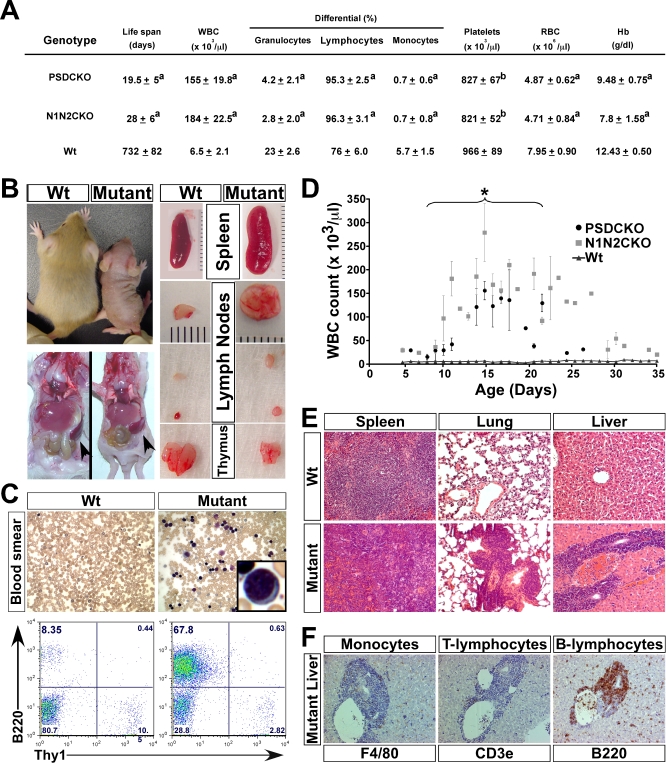
Figure 2. Mice Lacking Total Notch Signaling in the Skin Develop Severe B-LPD.
(A) Table shows the peripheral blood analysis of the mutant mice during the second week of life when WBC counts are at their maximum (n = 6, for each group). The values are presented as mean ± standard deviation (“a” indicates p < 0.001 and “b” indicates p < 0.05 (compared to wild-type control)).
(B) Macroscopic examination of a P14 mutant (N1N2CKO) compared to its wild-type littermate reveals the mutant's smaller body size yet significantly larger spleen and lymph nodes.
(C) Peripheral blood smear (Giemsa-stained; 250× magnification) and FC analysis show the appearance of lymphoblasts (inset) and the expansion of B220+ B cells in the mutant blood, respectively.
(D) Monitoring the mutant animals' WBC counts over their life span (n = 10 for each genotype) demonstrates a surge during the first 2 weeks and plateau during the third week of life, when most mutants die (asterisk). Note the trend of WBC counts toward normalization in a few mice that live a few days longer.
Severe B-LPD leads to infiltration of liver, lung, and spleen of the mutant animals with B220+ B cells shown on (E) hematoxylin-and-eosin-stained tissue sections and (F) antibody-stained liver sections (200× magnification).

