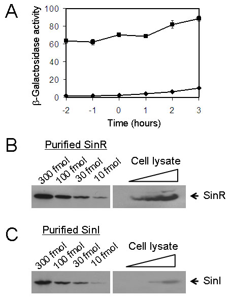
Figure 1. SinI levels greatly exceed that of SinR.

(A) Assays of β-galactosidase specific activity of cells carrying either the PsinR-lacZ (filled squares; strain YC108) or the PsinI-lacZ (filled diamonds; strain YC127) fusion at the amyE locus on the chromosome. Assays were performed for cells grown in MSgg medium and harvested at the indicated times. Time zero refers to the end of exponential phase growth. (B), (C) Quantitative immunoblots of SinR and SinI. Left-hand panels show affinity-purified, recombinant SinR and SinI proteins that were loaded at the indicated amounts. In the right-hand panels, cleared protein lysates prepared from early stationary phase cultures (one hour into stationary phase) were loaded on the same gel in a series of dilutions.
