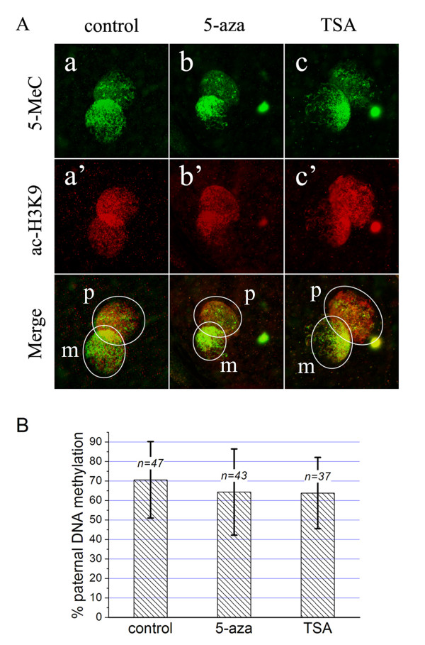
Figure 4.
Effects of 5-azacytidine or Trichostatin A on paternal demethylation. A. The oocytes were treated with 5-azacytidine (5-aza) or Trichostatin A (TSA) during the period of fertilization, and then they were double stained for DNA methylation (5MeC, green) and H3K9 acetylation (ac-H3K9, red). When compared to the untreated zygotes (control, a), the zygotes treated with 5-aza (b) or TSA (c) showed no significant changes in paternal DNA methylation levels. However, TSA treatment increased the immunoflurescence for ac-H3K9 (a', b', c'). In the merged images, the pronucleus area is marked by a white circle (p: paternal, m: maternal). B. Quantitative analysis of the DNA methylation levels in paternal pronucleus. The percentage of paternal DNA methylation indicates the DNA methylation levels in paternal pronucleus relative to maternal pronucleus (mean ± SD). The number of zygotes analyzed is indicated as "n=" above each column. Student's t test revealed no significant differences among groups (p > 0.05).

