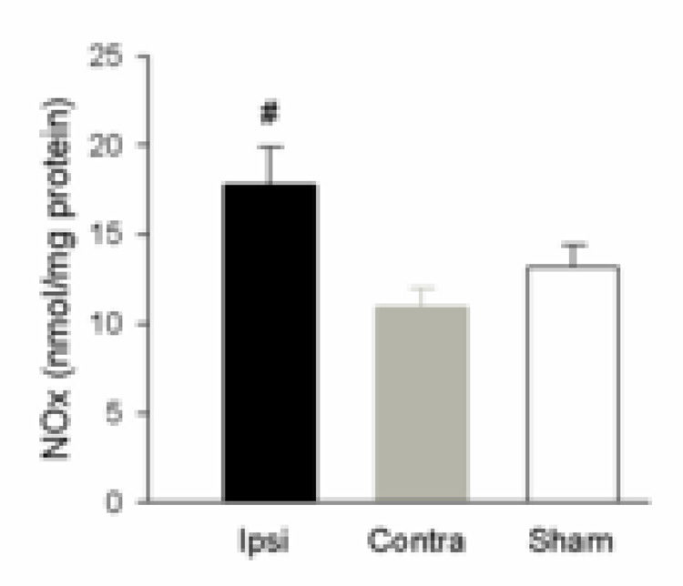
Figure 2.
Brain tissue NOx levels in cortex with MCAO. Brain tissue from ischemic cortex (Ipsi, n=7), contralateral cortex (Contra, n=7) and the cortex from animals that had sham surgery (Sham, n=4) was assayed for NOx, and normalized to protein. The level of NOx in tissue was determined in duplicate. NOx in ischemic cortex (Ipsi) was significantly higher than contralateral cortex (Contra) (#, P < 0.05).

