FIGURE 1.
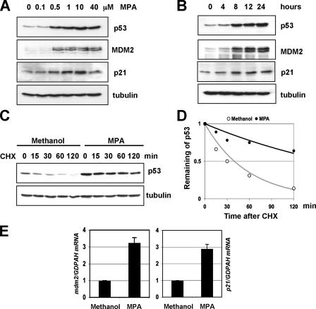
MPA treatment stabilizes and activates p53. A, dose-response of p53 induction and activation by MPA. U2OS cells were treated with different doses of MPA as indicated for 12 h. Cell lysates were assayed for expression of p53, p21, and MDM2 by immunoblot analysis. B, time-dependent effect of MPA on p53 induction and activation. U2OS cells were treated with 10 μmol/liter of MPA for different time courses as indicated. Cell lysates were assayed for expression of p53, p21, and MDM2 by immunoblot analysis. C and D, MPA treatment stabilizes p53. U2OS cells were treated with 10 μmol/liter MPA for 12 h, and then 50 μg/ml cycloheximide was added to the medium. The cells were harvested at different time points as indicated and assayed for levels of p53 and tubulin by immunoblot. The bands were quantified and normalized with loading controls determined by tubulin expression and plotted in D. E, MPA treatment induces the expression of p21 and mdm2 mRNA levels. U2OS cells were treated with methanol or 10 μmol/liter MPA for 12 h. Total RNAs were prepared from cells and retrotranscribed. Real-time PCR analysis was then conducted to determine the relative expression of the p21 and mdm2 mRNA as normalized against GAPDH mRNA.

