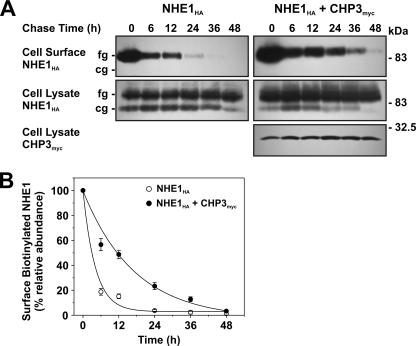
FIGURE 11.
Effect of CHP3 on half-life of cell surface NHE1 tagged by biotinylation. A, AP-1 cells stably expressing NHE1HA or stably coexpressing NHE1HA and CHP3myc were subject to cell surface biotinylation, as described under “Experimental Procedures.” The cells were returned to growth media at 37 °C, and then cell lysates were prepared at varying times over a 48-h period. At each time point, a small fraction of the cell lysates was removed for immunoblotting, and the remainder was incubated with 200 μl of NeutrAvidin-Sepharose beads to extract the biotinylated proteins. Total cellular levels of fully glycosylated (fg) and core-glycosylated (cg) NHE1HA and CHP3myc as well as levels of surface biotinylated, fully glycosylated NHE1HA were monitored as a function of time by SDS-PAGE and immunoblotting, as described in the legend to Fig. 6. B, data represent densitometric analysis of the cell surface biotinylated NHE1HA presented in A, normalized as a percentage of its maximal abundance at time 0 h. Values represent the mean ± S.E. of four experiments. Error bars smaller than the symbol are absent.

