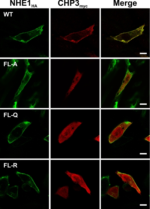
FIGURE 4.
Colocalization of NHE1 and CHP3 in intact cells. AP-1 cells were stably transfected with either wild-type (WT) or mutant forms (FL-A, -Q, or -R) of NHE1HA, followed by transient coexpression of CHP3myc, and their subcellular distribution was compared by immunofluorescence confocal microscopy. NHE1HA was detected with a primary mouse monoclonal anti-HA antibody and a secondary goat anti-mouse antibody conjugated to Alexa Fluor™ 488. CHP3myc was detected with a rabbit polyclonal anti-Myc antibody and a secondary goat anti-rabbit antibody conjugated to Alexa Fluor™ 568. The scale bar in the panels on the right represents 10 μm.

