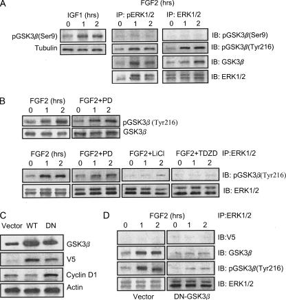
FIGURE 4.
FGF2-mediated association between ERK1/2 and GSK3β. A, SK-N-MC cells were treated with FGF2 (10 ng/ml) for the indicated times. Total protein lysates were collected and immunoprecipitated (IP) with either anti-ERK1/2 or anti-pERK1/2 antibody. The immunoprecipitates were then immunoblotted (IB) for the expression of GSK3β, pGSK3β(Ser-9 and Tyr-216), and ERK. IGF1-induced pGSK3β(Ser-9) served as a positive control. The experiment was replicated three times. B, SK-N-MC cells were pretreated with inhibitors of MEK1 (PD98059, 50 μm), JNK (JNK-I, 1 μm), p38 MAPK (SB20358, 10 μm), and GSK3β (LiCl, 20 mm; TDZD, 10 μm) for 30 min. After that, cells were exposed to FGF2 (10 ng/ml) for the indicated times. Cell lysates were immunoprecipitated with an anti-ERK1/2 antibody and immunoblotted using an anti-pGSK3β(Tyr-216) antibody. The experiment was replicated three times. C, SK-N-MC cells were transfected with either an empty vector or GSK3β constructs (wild type, WT; dominant negative, DN) for 48 h. The expression of GSK3β, V5 tag protein, cyclin D1, and actin was examined with immunoblotting. D, SK-N-MC cells were transfected with either an empty vector or a dominant negative GSK3β construct for 48 h. After that, cells were treated with FGF2 (10 ng/ml) for the indicated times. Cell lysates were immunoprecipitated with an anti-ERK1/2 antibody and immunoblotted for the expression of V5 tag protein, GSK3β, pGSK3β(Tyr-216), and ERK1/2. The expression was replicated three times.

