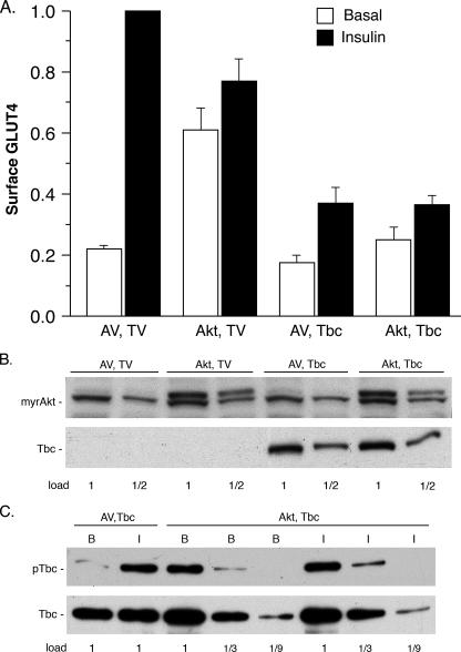
FIGURE 3.
Effect of myrAkt on cell surface GLUT4 and Tbc1d1 phosphorylation. A, 3T3-L1 adipocytes were co-transfected with the plasmid for HA-GLUT4-GFP and the plasmids for myrAkt1 with a carboxyl-terminal Myc tag (Akt) and 3× FLAG-tagged human Tbc1d1 (Tbc) or for the corresponding empty vectors (AV, TV). Cell surface HA-GLUT4-GFP was measured as described under “Experimental Procedures.” The values are the averages ± S.E. for four experiments. The values in each experiment were normalized to 1.0 for the AV, TV control in the insulin state. B, immunoblots of the SDS samples of the transfected cells in A for myrAkt with anti-Myc tag and for Tbc1d1 with anti-FLAG tag. The 1× load contained 20 μg of protein. The anti-Myc also cross-reacted with a protein that migrated just below myrAkt, which serves as a loading control. C, SDS/C12E9 lysates of the transfected cells in A were immunoprecipitated with anti-FLAG agarose. The immunoadsorbates were immunoblotted for phosphorylation of Tbc1d1 with the PAS antibody (upper panel) and for Tbc1d1 with anti-FLAG (lower panel). A repetition of B and C with a second set of samples gave similar results.

