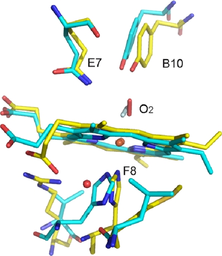
FIGURE 6.
Oxygen heme moiety. Least square superposition of HbIILp (yellow) and HbAsc (cyan, PDB code 1ASH). The superposition was carried out taking into account all the residues in the structure (amino acids and prosthetic group). The water molecule forming a hydrogen bond between the propionate group and His97(F8) in HbIILp is also shown. This water molecule is not present in the oxygen bound structure of HbAsc. The oxygen molecule shows opposite orientation in both structures (the oxygen molecule in HbAsc is shown in clear cyan).

