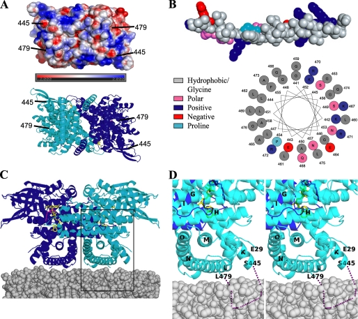
FIGURE 4.
Putative membrane binding site of VLCAD. A, an electrostatic representation (top) and a ribbon representation of the membrane-binding surface are shown (90° rotation from Fig. 1 along the x-axis). B, a helix model built from residues 441-481, which include the disordered residues. The helix shows a periodicity that is strikingly amphipathic in nature and may be capable of interacting with the mitochondrial membrane. C, putative VLCAD membrane orientation. It is proposed that residues 445-479 are responsible for anchoring VLCAD to the membrane in the orientation pictured. D, magnification and stereo view of the rectangular region of Fig. 4C. The dotted purple line represents the disordered region.

