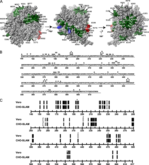
FIGURE 1.
Mutagenesis of predicted interaction regions of MV H. A, MV H model, with residues predicted to be part of protein-protein interactions depicted in red (SLAM-specific residues), blue (CD46-specific residues), and green (residues mutated in this study). The left panel is a view of the protein from the top; in the center panel the model was rotated 90° around the x-axis; the right panel was generated by rotating the center model 180° around the y-axis. The molecule has been depth-cued using the fog function to give perspective. B, MV H sequence and the mutations introduced. Single and multiple amino acid substitutions are indicated above the sequence. Alanine (A) was used to replace charged and polar residues and serine (S) was used to replace apolar residues. C, fusion efficiency of the H-protein mutants in Vero or CHO-SLAM cells. Rectangles represent multiple amino acid substitutions; bars represent single amino acid substitutions. Filled objects indicate full fusion activity, void objects indicate lack of fusion activity, and partially filled objects indicate intermediate fusion levels as defined under “Experimental Procedures.” Single amino acid substitutions that are SLAM-blind or CD46-blind are indicated by an asterisk (*) or a hash mark (#), respectively.

