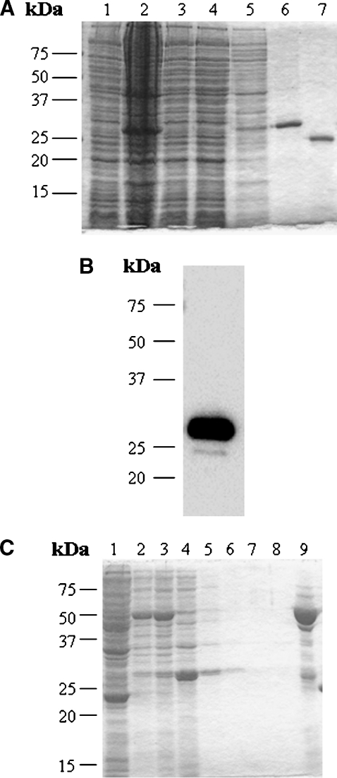
Fig. 1.
Purification and identification of recombinant human StarD4 protein. A: SDS-PAGE analysis and Coomassie blue staining of human StarD4 overexpressed in BL21 cells at each step of the purification. Lane 1, total soluble protein before induction (30 μg); lane 2, total soluble protein after induction (50 μg); lane 3, total soluble protein after incubation with the resin (30 μg); lane 4, proteins eluted with wash buffer I (30 μg); lane 5, proteins eluted with wash buffer II (10 μg); lane 6, protein eluted with lysis buffer plus 1 M imidazole (20 μg); lane 7, StarD4 protein after cleavage of the His tag with recombinant enterokinase. A major protein band was found around 27 kDa. B: Western blot analysis of human StarD4 with monoclonal anti-polyhistidine antibody. Protein eluted with lysis buffer plus 1 M imidazole (200 ng). C: SDS-PAGE analysis and Coomassie blue staining of GST-StarD4 fusion protein overexpressed in BL21 cells at each step of the purification. Lane 1, total soluble protein before induction (50 μg); lane 2, total soluble protein after induction (30 μg); lane 3, total soluble protein before incubation with Glutathione Sepharose 4B (30 μg); lane 4, proteins not bound by the resin (30 μg); lanes 5 to 8, proteins eluted with PBS (10 μg); lane 9, protein eluted with reduced glutathione (20 μg).

