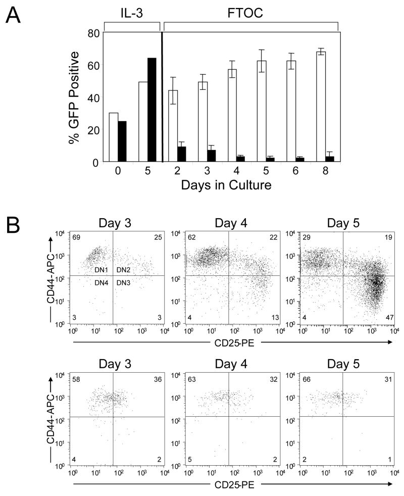
Fig 2.
TLX1 blocks thymocyte differentiation at the DN stage. (A) Time course analysis of TLX1-dependent thymocyte arrest. FL precursors transduced with either MSCV-GW (white bars) or TLX1 (black bars) were used in FTOCs. Aliquots of transduced FL precursors were maintained in parallel in liquid cultures containing IL-3. Thymocytes from organ cultures were analyzed at indicated time points for expression of GFP. Data represent the mean ± SEM for 3–4 lobes analyzed per time point. (B) Analysis of DN differentiation of FL precursors transduced with MSCV-GW (upper plots) or TLX1 (lower plots) retroviral vectors. Thymocytes from FTOCs were processed on days 3, 4 and 5 of culture and immunophenotyped for analysis of CD44/CD25 populations by gating on GFP+CD4−CD8− cells. Bivariate histograms are representative of 6–8 thymic lobes from two independent experiments.

