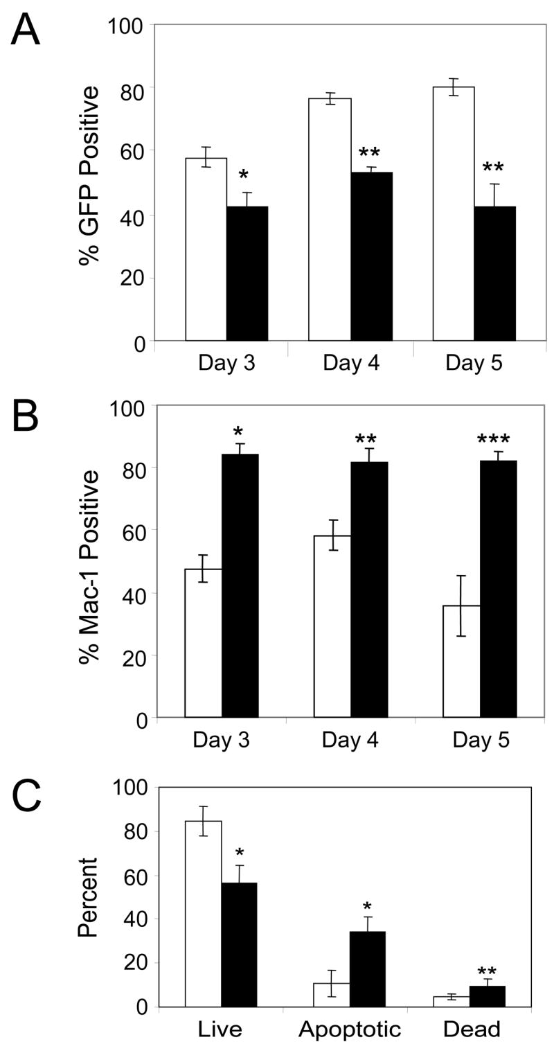
Fig 3.

Decreased numbers of thymocytes in TLX1 FTOCs. (A) GFP expression in thymocytes from days 3, 4 and 5 of FTOCs. Data represent the mean ± SEM of 8–10 fetal thymi per time point from two independent experiments for MSCV-GW (white bars) and TLX1 (black bars); * P < 0.003, ** P < 0.0001. (B) MSCV-GW- (white bars) or TLX1- (black bars) expressing thymocytes were analyzed at FTOC days 3, 4 and 5 for surface expression of the FL stem cell/myeloid marker Mac-1. Data are based on a GFP+ gate and represent the mean ± SEM of 6 lobes; * P < 0.0002, ** P < 0.011, ***P < 0.0001. (C) Viability of thymocytes in day 5 FTOCs. MSCV-GW- (white bars) and TLX1 retroviral vector- (black bars) transduced thymocytes were stained with 7-AAD. Cells were gated on GFP fluorescence and the proportions of 7-AAD negative (live), dim (early apoptotic) and bright (late apoptotic or necrotic, dead) cells were determined. Data represent the mean ± SEM of at least four lobes per vector; * P < 0.0016, ** P < 0.036.
