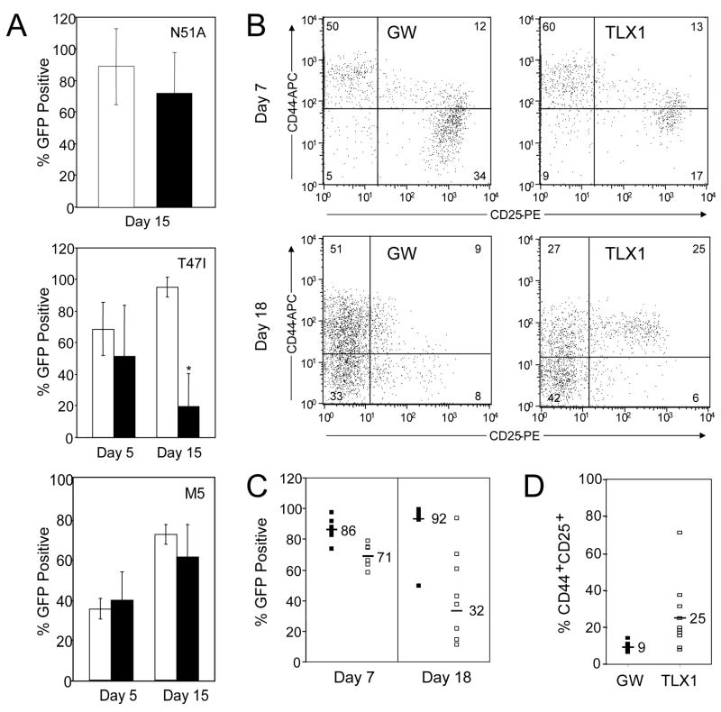
Fig 5.
Mechanistic aspects of TLX1 inhibition of thymocyte differentiation. (A) Disruption of residues implicated in DNA binding and PBX cofactor interactions abrogates TLX1-mediated differentiation block. Fetal thymocytes transduced with TLX1 N51A, TLX1 T47I and TLX1 M5 (black bars) were analyzed from day 5 and/or day 15 FTOCs for GFP expression versus MSCV-GW (white bars). Graphs present the mean ± SEM of data from 7–14 lobes per retroviral vector and 2–3 independent experiments; *P < 0.0001. (B–D) Transgenic BCL2 partially overcomes TLX1-inhibited thymic repopulation. (B) Phenotype of BCL2-expressing DN thymocytes at day 7 of FTOCs transduced with MSCV-GW or TLX1 retroviral vectors. Representative bivariate histograms of CD44 versus CD25 gated on GFP+/CD4-CD8- cells. (C) GFP expression in day 7 (MSCV-GW: N = 6; TLX1: N = 6) and day 18 (MSCV-GW: N = 12; TLX1: N = 8) BCL2-expressing thymocytes. MSCV-GW (black bars) and TLX1 (white bars). Data shown were combined from two independent experiments on day 18 and the means for the data subsets are indicated. Day 18 percentage of TLX1+GFP+ cells is significantly different; P < 0.0001. (D) Percentage of CD44+CD25+ cells is increased in TLX1+ BCL2-expressing thymocytes compared with MSCV-GW cells analyzed at day 18. MSCV-GW: N = 12; TLX1: N = 10. Means of the subsets are indicated; P < 0.0076.

