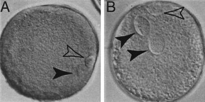
Figure 1.
Bovine oocytes matured in vitro demonstrate distinctive nuclear configuration compared with zygotes at the pronuclear stage. Oocytes were fixed 18 hr after placing in maturation medium and zygotes were fixed 18 hr postsemen addition, in three parts ethanol/one part acetic acid for 24 hr, before acid-orcein staining (1% orcein stain in 40% acetic acid in H2O). (A) After induction of maturation, the oocyte undergoes germinal vesicle (4N) breakdown, the chromatin condenses, the first meiotic division occurs, and the first polar body, containing half of the genome (2N), is extruded. The remaining condensed, diploid chromatin is aligned at the second metaphase plate (arrowhead). Chromatin is not enclosed in a nuclear membrane and is located next to the first polar body. The polar body also has intense chromatin, which is enclosed in a plasma membrane during autosome segregation (open arrowhead). The oocyte remains arrested in MII until the completion of meiosis, which is heralded by the extrusion of the second polar body (1N) induced by fertilization. (B) After fertilization, the oocyte progresses from MII to interphase. Zygote contains both maternal and paternal pronuclei enclosed by a nuclear envelope (arrowhead). The second polar body is located next to the maternal pronucleus with distinctive chromatin staining (open arrowhead). In addition to the difference in nuclear configuration, oocytes have very condensed chromatin and intense chromatin staining, whereas zygotes have dispersed chromatin with less intense staining.

