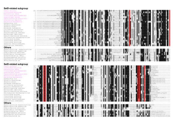
Figure 3.
Multiple alignment of hypothetical protein HP1. Representative sequences were divided into two groups: SelD-related subgroup and other homologs. Residues which are strictly conserved in the SelD-related subgroup are shown in red background. Other residues shown in white on black or grey are conserved in homologs. Organisms containing orphan SelD are highlighted in pink font.

