
Fig. 1.
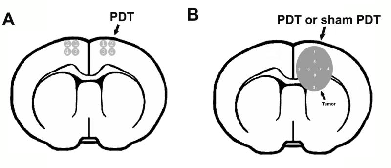
Four high magnification fields from digitized images of the PDT-treated region and corresponding contralateral area were selected for immunohistochemistry analysis (A). Eight high magnification fields from digitized images of tumor implanted in nude mice with/without pro-PDT treatment (B).