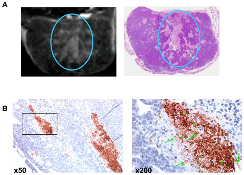
Fig. 4.

A: Representative T1-weighted MRI image from a mouse bearing a subcutaneously inoculated Colo-205 tumor (left) and a photograph of the corresponding H&E-stained section of the tumor (right). The mouse received intravenous injections of L-PG-DTPA-Gd (0.04 mmol Gd/kg). L-PG-DTPA-Gd was distributed within the necrotic area (circle). B: Co-localization of macrophages and biotinylated L-PG-DTPA-Gd in the necrotic area in OCA-1 tumors. Staining with antibody directed against macrophage antigen F4/80 (reddish-pink) and with streptavidin-horseradish peroxidase and DAB for biotinylated L-PG-DTPA-Gd showed widespread macrophage infiltration in necrotic areas as well as distribution of the polymer and its degradation products to the necrotic area (brown). The sections were counterstained with hematoxylin (blue) to label the nuclei of viable tumor cells. Examples of biotin-L-PG-DTPA-Gd staining and dual F4/80 and biotin staining are indicated with arrows and arrowheads, respectively (from reference [20].
