
Fig. 6.
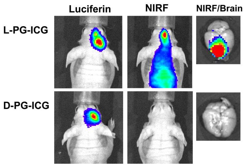
Representative in vivo near-infrared fluorescence (NIRF) images of cathepsin B activity in intracranially inoculated U87/TGL tumors. The mice used in the study had the same tumor burden, as indicated by the bioluminescence signal generated after intravenous injection of luciferin. NIRF Images were acquired 24 h after intravenous injection of L-PG-NIR813 (50 nmol/mouse) or D-PG-NIR813 (50 nmol/mouse) with the same NIR dye loading (10%) and the same molecular weight (17,500). The NIRF signal was visualized in the mouse injected with L-PG-NIR813 but not the mouse injected with D-PG-NIR813 (from reference [25]).