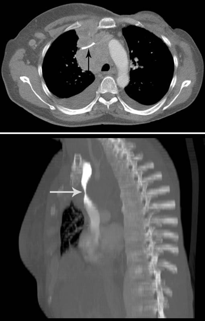
Fig 1 Contrast enhanced computed tomography in October 2004 shows enlarged mediastinal nodes with significant focal narrowing of the superior vena cava (top; arrow). Contrast enhanced computed tomography, with reconstruction using maximum intensity projection in the sagittal plane, shows focal narrowing of the superior vena cava (bottom; arrow)
