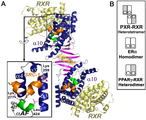
Figure 1. Structural Features of the PXR-RXR Heterotetramer.
(A) A model of the PXR (blue, magenta, green)-RXR (yellow) heterotetramer highlights the PXR homodimer interface and the ten-stranded intermolecule β-sheet formed between the two monomers. PXR residues Trp-223 and Tyr-225 central to homodimerization are rendered in yellow with transparent CPK spheres. The α-helices 3, 3′, 4 and αAF (green) create the AF-2 surfaces that bind leucine-rich coactivator peptides like SRC-1 (orange) using a charge clamp (Lys-259, Glu-427) and other residues (light pink). (B) Schematics of the oligomeric NR complexes examined in this paper.

