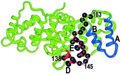
Figure 1.
Crystal structure of hydra annexin XII (2). Helices A and B (domain 3) and helices D and E (domain 2) are highlighted in blue and red, respectively. Sites of the spin-labeled residues 138–163 are indicated by residue labels and solid spheres on the α-carbons.

