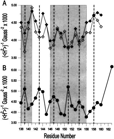
Figure 4.
Inverse second moment (〈H2〉) for the EPR spectra of R1 as a function of sequence position. (A) Annexin XII in solution at pH 7.4 (solid line) and at pH 4.0 (dashed line). (B) Annexin XII bound to vesicles at pH 4.0 in the absence of Ca2+. The gray boxes on the left and right correspond to residues in the D and E helices, respectively, in the crystal structure of annexin XII (2).

