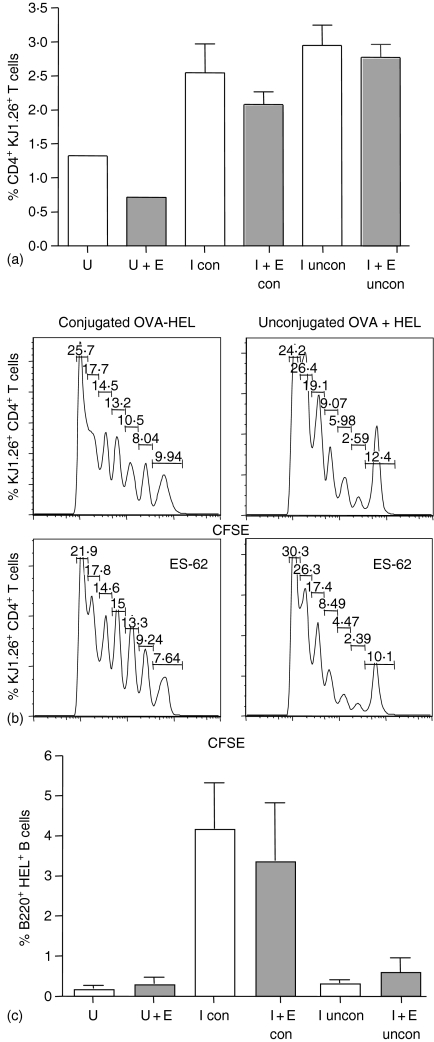
Figure 5.
Effects of ES-62 on antigen-specific CD4+ T-cell and B220+ B-cell clonal expansion in vivo following immunization with unconjugated OVA and HEL. Non-transgenic BALB/c recipient control and ES-62-treated mice were injected with 2·5 × 106 CD4+ KJ1.26+ T cells and B220+ HEL+ B cells. Twenty-four hr later, these mice were either immunized s.c. with 130 µg of conjugated OVA–HEL (con) or unconjugated OVA and HEL (uncon) in CFA. The percentage of CD4+ KJ1.26+ T cells (a) and B220+ HEL+ B cells (c) in the draining lymph nodes of unimmunized control (U) and unimmunized ES-62-treated (U + E) and immunized control (I) and immunized ES-62-treated (I + E) adoptively transferred recipients was assessed by flow cytometry on day 7 after immunization. Data are presented as a bar chart of the mean ± SEM for three mice per group. Prior to transfer, T cells were labelled with CFSE and five days after immunization, draining lymph nodes from immunized control and ES-62-treated adoptive transfer recipients were removed and CFSE fluorescence, a measure of cell division, was assessed in CD4+ KJ1.26+ T cells by flow cytometry (b) Data are shown as histograms of CFSE fluorescence versus number of CD4+ KJ1.26+ T cells and are from one mouse, which is representative of three mice per group and a total of two independent experiments.

