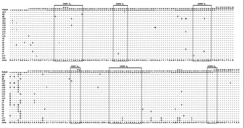
Figure 2.
Alignment of amino acid sequences of related scFvs binding to GCN4(7P14P) peptide, isolated by ribosome display. Only VL and VH are shown. Residues identical to the consensus sequence of the 22 related scFvs (cons) are represented by dashes. The upper three scFvs (g2, g5, and g14) were isolated in experiment B, and the other 19 scFvs (c1 to c23) in experiment A (see Fig. 1). Numbering of amino acid residues in VL and VH and the labeling of CDRs is according to Kabat et al. (23).

