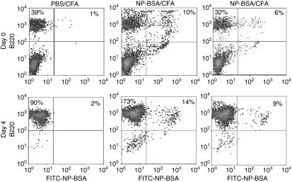
Figure 4.
Growth of antigen-specific B cells after CD40L activation. Mice were injected three times with PBS/CFA or NP-BSA/CFA. Ten days after the last treatment, mice were killed and an aliquot of spleen cells stained with PE–anti-B220 antibody and with FITC–conjugated NP-BSA. The remainder of the spleen cells were cultured on CD40L-transfected L cells in the presence of rIL-4 and cyclosporin A for 4 days and phenotyped. Representative data from a total of 9 mice on day 0 and 6 mice on day 4 are presented.

